
POCUS cardiac: Parasternal long axis view with pericardial effusion tracking above the descending aorta and RV diastolic collapse.
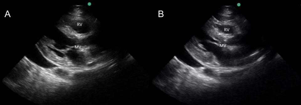
The most specific sonographic finding for cardiac tamponade is RV diastolic collapse. Diastole corresponds with the opening of the mitral valve. While the mitral valve is open you can see that the RV is collapsing (still B). Still image A above corresponds with systole.
A middle aged female with a history of HIV presents with progressive leg swelling for 2 months. She is hypertensive (200/150), tachycardic, and with good oxygen saturation on room air. Patient is in no respiratory distress but has bilateral lower extremity edema extending proximally to the thighs, sacrum, and the abdominal wall.
POCUS Cardiac: The parasternal long axis view shows a pericardial effusion and diastolic right ventricular (RV) collapse, concerning for cardiac tamponade. Patient is admitted to the cardiac intensive care unit where a pericardiocentesis drained a total of 1000 ml of serosanguinous fluid over the course of the evening.
POCUS pearl: The most specific sonographic finding for cardiac tamponade is RV diastolic collapse. Diastole corresponds with the opening of the mitral valves (still image B above).